a) Levels of organization
2.1: Describe the levels of organization within organisms: organelles, cells, tissues, organs and systems
- Organelles are highly organized structures of molecules that have a specific function within a cell (e.g. mitochondria)
- Cells are made up of organelles and are the building blocks of life (e.g. palisade cells)
- Tissues are a collection of similar cells performing a common function (e.g. muscle tissue)
- Organs are made up of several kinds of tissues to make a functional unit (e.g. heart)
- Systems are made up of several organs (e.g. respiratory system)
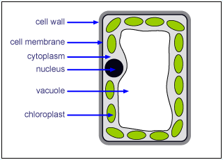
b) Cell structure
2.2: Recognize cell structures, including the nucleus, cytoplasm, cell membrane, cell wall, chloroplast and vacuole
|
2.3: Describe the functions of the nucleus, cytoplasm, cell membrane, cell wall, chloroplast and vacuole
Nucleus • Contains DNA • Controls cell operations Cytoplasm • Cell reactions occur here • Contains vital organelles Cell Membrane • Controls what enters and exits the cell • Provides structural support Cell Wall • Provides structural support • Made of cellulose Chloroplast • Contains chlorophyll where photosynthesis occurs Vacuole • Storage of water and cell sap |

|
2.4: Describe the differences between plant and animal cells
- Plant cells are larger than animal cells
- Plant cells contain chloroplasts
- Plant cells have conspicuous vacuoles
- Animal cells can engulf other cells
c) Biological molecules
2.5: Recall the chemical elements present in carbohydrates, proteins, and lipids (fats and oils)
Carbohydrates
• Oxygen
• Hydrogen
• Carbon
Proteins
• Oxygen
• Hydrogen
• Carbon
• Nitrogen
Fats
• Oxygen
• Hydrogen
• Carbon
Carbohydrates
• Oxygen
• Hydrogen
• Carbon
Proteins
• Oxygen
• Hydrogen
• Carbon
• Nitrogen
Fats
• Oxygen
• Hydrogen
• Carbon
2.6: Describe the structure of carbohydrates, proteins and lipids as large molecules made up from smaller basic units
Carbohydrates
• Starch
• Cellulose
• Glycogen
Proteins
• Amino acids
Fats
• Fatty acids
• Glycerol
Carbohydrates
• Starch
• Cellulose
• Glycogen
Proteins
• Amino acids
Fats
• Fatty acids
• Glycerol
2.7: Describe the tests for glucose and starch
Starch Test
Starch Test
- Take leaf
- Dip in boiling water for one minute
- Place leaf in boiling tube containing ethanol
- Place boiling tube in hot water bath for ten minutes
- Transfer leaf to white tile and add iodine solution; parts of leaf with starch present will turn dark blue or black
- Repeat
|
Glucose Test
|
"Girls think Benedict Cumberbatch is sweet. Like glucose."
- Matthew Tan Benedict solution is used to test for glucose. |
2.8: Understand the role of enzymes as biological catalysts in metabolic reactions
Enzymes are soluble protein molecules that can speed up reactions in cells without being used up themselves
Enzymes are soluble protein molecules that can speed up reactions in cells without being used up themselves
2.9: Understand how the functioning of enzymes can be affected by changes in temperature
The warmer the enzyme environment is, the more efficiently it will work. This applies until ~37°C when the enzyme denatures (completely stops working).
The warmer the enzyme environment is, the more efficiently it will work. This applies until ~37°C when the enzyme denatures (completely stops working).
2.10: Understand how the functioning of enzymes can be affected by changes in pH
As pH gets higher, enzyme activity gets more efficient until the optimum pH is reached. After that, the enzyme activity gets less efficient
As pH gets higher, enzyme activity gets more efficient until the optimum pH is reached. After that, the enzyme activity gets less efficient
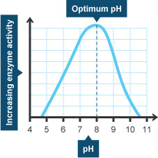
There are different optimum pH levels for different enzymes:
Lipase (Pancreas) - pH 8
Lipase (Stomach) - pH 4 - 5
Pepsin - pH 1.5 - 1.6
Amylase (Pancreas) - pH 6.7 - 7
Catalase - pH 7
Lipase (Pancreas) - pH 8
Lipase (Stomach) - pH 4 - 5
Pepsin - pH 1.5 - 1.6
Amylase (Pancreas) - pH 6.7 - 7
Catalase - pH 7
2.11: Describe how to carry out simple controlled experiments to illustrate how enzyme activity can be affected by changes in temperature
- Cut out a strip of potato using a cork borer. Chop this into pieces which are 5mm in width. Add these pieces to a test tube and put the test tube into a 21°C water bath
- Add 5ml of hydrogen peroxide solution to the first tube and start the stopwatch
- Time two minutes and then measure the height of the foam in mm
- Repeat but vary the temperatures (e.g. 21°C, 25°C, 30°C, 35°C, 40°C)
- Repeat the entire experiment and average out the results
d) Movement of substances into and out of cells
|
2.12: Recall simple definitions of diffusion, osmosis and active transport
Diffusion: When air particles move from an area of higher concentration to an area of lower concentration until they are evenly spread out Osmosis: When water particles move from an area of higher concentration to an area of lower concentration until they are evenly spread out Active Transport: When some particles move against the flow of others (lower concentration to higher concentration) |

|
2.13: Understand that the movement of substances into and out of cells can be by diffusion, osmosis and active transport
Diffusion: Oxygen diffuses into the human lungs and into the blood
Osmosis: Water osmoses through root cells in plants
Active Transport: Requires energy from respiration (e.g. active transport from small intestine to blood)
Diffusion: Oxygen diffuses into the human lungs and into the blood
Osmosis: Water osmoses through root cells in plants
Active Transport: Requires energy from respiration (e.g. active transport from small intestine to blood)
2.14: Understand the importance of turgid cells in plants as a means of support
Turgid cells are cells in which the vacuole contains the maximum amount of water possible. This water enters the plant cell via osmosis. The plant cell becomes hard and has a rigid shape that can't be changed. This helps support the plant.
Turgid cells are cells in which the vacuole contains the maximum amount of water possible. This water enters the plant cell via osmosis. The plant cell becomes hard and has a rigid shape that can't be changed. This helps support the plant.
2.15: Understand the factors that affect the rate of movement and substances into and out of cells including the effects of surface area to volume ratio, temperature, and concentration gradient
Surface Area to Volume Ratio
The higher the surface area to volume ratio, the faster the rate of diffusion
Temperature
The higher the temperature, the faster the rate of diffusion
Concentration Gradient
The higher the concentration gradient, the faster the rate of diffusion
Surface Area to Volume Ratio
The higher the surface area to volume ratio, the faster the rate of diffusion
Temperature
The higher the temperature, the faster the rate of diffusion
Concentration Gradient
The higher the concentration gradient, the faster the rate of diffusion
2.16: Describe simple experiments on diffusion and osmosis using living and non-living systems
Diffusion
Diffusion
- Put a colored substance (like food coloring) into water
- Observe the colored substance make the water the same color
- Get two near-identical pieces of potato
- Put one in distilled water and one in salt water
- Weigh them both after a given time
- The one from the salt water will have lost mass, whereas the other one will have gained mass
e) Nutrition
|
2.17: Describe the process of photosynthesis and and understand its importance in the conversion of light energy to chemical energy
|

|
2.18: Recall the word equation and the balanced chemical equation for photosynthesis
Word Equation
Carbon Dioxide + Water + (Sunlight) → Glucose + Oxygen
Chemical Equation
6CO2 + 6H2O + (Sunlight) → C6H12O6 + 6O2
Word Equation
Carbon Dioxide + Water + (Sunlight) → Glucose + Oxygen
Chemical Equation
6CO2 + 6H2O + (Sunlight) → C6H12O6 + 6O2
2.19: Understand how carbon dioxide concentration, light intensity and temperature effect the rate of photosynthesis
Carbon Dioxide
The higher the carbon dioxide concentration, the faster the rate of photosynthesis
Light Intensity
The higher the light intensity, the faster the rate of photosynthesis
Temperature
The higher the temperature, the faster the rate of photosynthesis (unless the temperature gets high enough to kill the plant)
2.20: Explain how the structure of the leaf is adapted for photosynthesis
- The leaf has a large surface area to volume ratio which means it contains the maximum amount of chlorophyll possible, so they can absorb the maximum possible amount of sunlight to aid photosynthesis
- The waxy cuticle on the top of the leaf is transparent which allows light to pass through
- Palisade cells are packed with chloroplasts and they are near the top of the leaf
- Spongy cells have air spaces between them, allowing CO2 to diffuse through them easily
2.21: Recall that plants require mineral ions for growth and that magnesium ions are needed for chlorophyll and nitrate ions are needed for amino acids
- Mineral ions are needed for plant growth
- Magnesium ions are needed for chlorophyll
- Nitrate ions are needed for amino acids
2.22: Describe simple controlled experiments to investigate photosynthesis, showing the evolution of oxygen from a water plant, the production of starch and the requirements of light, carbon dioxide, and chlorophyll
- Place pond weed under water
- Vary the factors:
- Move a lamp further away from the plant
- Add baking powder to the water (it increases carbon dioxide levels)
- Test a white leaved plant and a green leaved plant (the green plant has more chlorophyll)
- Use iodine solution to test for starch (see 2.7)
2.23: Understand that a balanced diet should include appropriate portions of different dietary components
- Carbohydrates
- Proteins
- Lipids
- Vitamin A
- Vitamin C
- Vitamin D
- Dietary Fiber
- Calcium
- Iron
- Water
2.24: Recall sources and describe functions of different dietary components
- Dietary component: sources - functions - deficiency disease
- Carbohydrates: bread, pasta, cereal - production of energy - obesity (too many carbohydrates)
- Proteins: animal products - building block for muscle, skin, hair, and other tissues - kwashiorkor (not enough proteins)
- Lipids: dairy products - preserve body heat and protect organs - heart disease (too many lipids)
- Vitamin A: vegetables, carrots - growth of skin cells and vision in dim light - night blindness, damaged cornea (not enough Vitamin A)
- Vitamin C: strawberries, oranges - formation of teeth, gums and blood vessels - scurvy (not enough Vitamin C)
- Vitamin D: sunlight - absorption and use of calcium and phosphate for healthy bones and teeth - rickets, poor teeth (not enough Vitamin D)
- Dietary Fiber: cereal - maintaining normal cholesterol and blood sugar levels - diabetes, obesity, heart disease (not enough dietary fiber)
- Calcium: dairy products - growing bones and teeth - osteoporosis (not enough calcium)
- Iron: meats, beans, apples, iron supplement - transportation of oxygen - anaemia (not enough iron)
- Water: water - digestion, maintaining body temperature, removal of waste by urine - dehydration (not enough water)
2.25: Understand that energy requirements vary with activity levels, age and pregnancy
The energy requirements of a person vary with activity level, pregnancy, and age. For example, a blue-collar worker will definitely need more energy than a white-collar worker. A pregnant woman will also need more energy because she has to use lots of it on her child.
The energy requirements of a person vary with activity level, pregnancy, and age. For example, a blue-collar worker will definitely need more energy than a white-collar worker. A pregnant woman will also need more energy because she has to use lots of it on her child.
|
2.26: Recognize the structures of the human alimentary canal and describe in outline the features of the mouth, oesophagus, stomach, small intestine, large intestine, and pancreas
Mouth: The digestion process begins - the food is reduced in size until it is small enough to pass through the oesophagus Oesophagus: The food particles are transported into the digestive system Stomach: The acids in the stomach break the food down into a liquid mixture - gastric juices and stomach muscles grind and smash the food Small Intestine: Food is broken down so that it can be passed into the bloodstream. Villi in the small intestine absorb the nutrients - the nutrients diffuse into the villi Large Intestine: The liquid and nutrients are absorbed from the food particles and dry lumps of waste are left over Pancreas: The pancreas secrets enzymes that aid the process of digestion |

|
2.27: Understand the processes of ingestion, digestion, absorption, assimilation, and egestion
Ingestion: The process of food entering the body
Digestion: The mechanical and chemical processes of breaking down food molecules
Absorption: The process of nutrients and liquids entering the bloodstream from the small intestine
Assimilation: The build-up of food particles into useful molecules (e.g. the build up of proteins into muscles)
Egestion: The process of eliminating feces from the body via the anus
2.28: Explain how and why food is moved through the gut by peristalsis
Peristalsis involves involuntary waves of muscle contraction that keep food moving along in one direction through the digestive system. It is a chain reaction of pushing and pulling. Muscles in the gut contract and relax and thus push the mushy, smooth food bolus down the gut so it can be digested further and important substances needed for the body's functioning can be absorbed out of it.
Peristalsis involves involuntary waves of muscle contraction that keep food moving along in one direction through the digestive system. It is a chain reaction of pushing and pulling. Muscles in the gut contract and relax and thus push the mushy, smooth food bolus down the gut so it can be digested further and important substances needed for the body's functioning can be absorbed out of it.
2.29: Understand the role of digestive enzymes to include the digestion of starch to glucose by amylase and maltase, he the digestion of proteins to amino acids by proteases and the digestion of lipids to fatty acids and glycerol by lipases
Digestive enzymes are used to aid digestion.
Digestive enzymes are used to aid digestion.
- Amylase and maltase are used to digest starch into glucose
- Proteases are used to digest proteins into amino acids
- Lipases are used to digest lipids into fatty acids and glycerol
2.30: Recall that bile is produced by the liver and stored in the gall bladder, and understand the role of bile in neutralizing stomach acids and emulsifying lipids
Bile is made in the liver and stored in the gall bladder. It helps to break down lipids and also helps get rid of toxins in the liver. It is a very powerful antioxidant and can be used to neutralize stomach acids.
Bile is made in the liver and stored in the gall bladder. It helps to break down lipids and also helps get rid of toxins in the liver. It is a very powerful antioxidant and can be used to neutralize stomach acids.
2.31: Explain how the structure of a villus helps absorption of the products of digestion in the small intestine
- Villi have capillary beds inside them which allows the food to pass directly to the bloodstream
- Villi have an epithelial layer which is covered in microvilli
- Microvilli increase surface area allowing for maximum absorption potential
2.32: Recall how to carry out a simple experiment to determine the energy content in a food sample
- Measure the mass of the food sample
- Ignite the food sample
- Heat up the water using the ignited food sample as a heat source
- Measure the temperature rise of the water after the food has finished burning
- Measure the mass of the food after it has finished burning
- Calculate how much energy there was in the food sample
- Repeat
f) Respiration
2.33: Recall that the process of respiration releases energy in living organisms
Aerobic Respiration
Glucose + Oxygen → Carbon Dioxide + Water
Anaerobic Respiration
Glucose → Energy + Lactic Acid
Aerobic Respiration
Glucose + Oxygen → Carbon Dioxide + Water
Anaerobic Respiration
Glucose → Energy + Lactic Acid
2.34: Describe the differences between aerobic and anaerobic respiration
- Aerobic respiration requires oxygen to take place; anaerobic only requires glucose
- Aerobic respiration produces carbon dioxide and water as by-products; anaerobic only produces lactic acid
2.35: Recall the word equation and balanced symbol equation for aerobic respiration in living organisms
Glucose + Oxygen → Carbon Dioxide + Water + (Energy)
C6H12O6 + 6O2 → 6CO2 + 6H2O + (Energy)
Glucose + Oxygen → Carbon Dioxide + Water + (Energy)
C6H12O6 + 6O2 → 6CO2 + 6H2O + (Energy)
2.36: Recall the word equation for anaerobic respiration in plants and animals
Plants: Glucose → Ethanol + Carbon Dioxide (+ some energy)
Animals: Glucose → Lactic acid (+ some energy)
Plants: Glucose → Ethanol + Carbon Dioxide (+ some energy)
Animals: Glucose → Lactic acid (+ some energy)
2.37: Describe simple controlled experiments to demonstrate the evolution of carbon dioxide and heat from respiring seeds or other suitable living organisms
To demonstrate the evolution of carbon dioxide
1. Put the seeds into a sealed controlled environment that allows respiration to occur
2. Use hydrogen-carbonate indicator to detect the CO2 levels
3. Wait for 24 hours
4. The hydrogen-carbonate indicator will have turned purple due to the decrease in CO2
To demonstrate the evolution of heat
1. Put the seeds into a sealed controlled environment that allows respiration to occur
2. Record the initial temperature
3. Wait 24 hours and record the final temperature - you will notice an increase
g) Gas exchange
2.38: Understand the role of diffusion in gas exchange
Concentration of gases in our body is less than the concentration of gases outside therefore the air will move to the area of lower concentration (our body) until it is evenly spread out.
Concentration of gases in our body is less than the concentration of gases outside therefore the air will move to the area of lower concentration (our body) until it is evenly spread out.
2.39: Understand gas exchange (of carbon dioxide and oxygen) in relation to respiration and photosynthesis
In Respiration: Glucose + Oxygen → Carbon Dioxide + Water
Oxygen diffuses into the plant and carbon dioxide diffuses out
In Photosynthesis: Carbon Dioxide + Water → Glucose + Oxygen
Carbon dioxide diffuses into the plant and oxygen diffuses out
In Respiration: Glucose + Oxygen → Carbon Dioxide + Water
Oxygen diffuses into the plant and carbon dioxide diffuses out
In Photosynthesis: Carbon Dioxide + Water → Glucose + Oxygen
Carbon dioxide diffuses into the plant and oxygen diffuses out
2.40: Understand that respiration continues during the day and night, but that the net exchange of carbon dioxide and oxygen depends on the intensity of light
Respiration takes place during the day and night, but gas exchange depends on the light intensity: more gas exchange occurs when the light intensity is higher during the day.
Respiration takes place during the day and night, but gas exchange depends on the light intensity: more gas exchange occurs when the light intensity is higher during the day.
2.41: Explain how the structure of the leaf is adapted for gas exchange
• Leaves are thin which allows CO2 to diffuse through easily
• There are air spaces between the spongy cells which allows CO2 to diffuse through
• Leaves are thin which allows CO2 to diffuse through easily
• There are air spaces between the spongy cells which allows CO2 to diffuse through
2.42: Describe the role of the stomata in gas exchange
Guard cells control the size of the stomata opening. An open stomata allows carbon dioxide diffuse between the leaves and the atmosphere.
Guard cells control the size of the stomata opening. An open stomata allows carbon dioxide diffuse between the leaves and the atmosphere.
2.43: Describe simple controlled experiments to investigate the effect of light on net gas exchange from a leaf, using hydrogen-carbonate indicator
Hydrogen-carbonate indicator detects carbon dioxide - in a normal (0.04%) concentration it is orange. In an increase (>0.04%) it is yellow, and in a decrease (<0.04%) it turns purple. Put hydrogen-carbonate indicator in a sealed flask with a leaf and observe for a decrease.
Hydrogen-carbonate indicator detects carbon dioxide - in a normal (0.04%) concentration it is orange. In an increase (>0.04%) it is yellow, and in a decrease (<0.04%) it turns purple. Put hydrogen-carbonate indicator in a sealed flask with a leaf and observe for a decrease.
2.44: Describe the structure of the thorax, including the ribs, intercostal muscles, diaphragm, trachea, bronchi, bronchioles, alveoli, and pleural membranes
Ribs: 24 curved ribs make up the ribcage that protects several vital organs including the heart and lungs. This protected area of the chest is known as the thorax.
Intercostal Muscles: Intercostal muscles are muscles that are in between the ribs in the ribcage. They contain three layers: the external layer that helps with inhalation of air, the internal layer which helps with exhalation of air, and the bottom layer.
Diaphragm: The diaphragm is a dome-shaped piece of muscle. It is located at the bottom of the chest and is attached to the ribs.
Trachea: The trachea is made of cartilage and divides into two bronchi when it reaches the lungs. Each bronchus goes into each lung.
Bronchi: Bronchi are two smaller tubes that come out of the trachea when it reaches the lungs. There is a left bronchus and a right bronchus, both of which go into each lung and branch out into smaller bronchioles.
Bronchioles: Bronchioles are even smaller passages that begin at the end of the bronchi. The bronchioles fan out into the lungs and end in alveoli. Unlike bronchi, which have cartilage, bronchioles are lined with muscular walls. The muscular lining constricts the airways and controls the flow of air to the alveoli.
Pleural Membranes: The pleural membrane is a membrane found in the chest. It acts as a lubricant that lines the outside of the lungs: it allows minimum friction between the lung walls and the thoracic wall between breathing.
2.45: Understand the role of the intercostal muscles and the diaphragm in ventilation
Breathing in
• The intercostal muscles contract
• The diaphragm contracts and moves down
• These actions increase space inside the lungs, allowing air inside
Breathing out
• The intercostal muscles relax
• The diaphragm relaxes and moves up
• These actions decrease space inside the lungs, pushing air outside
2.46: Explain how alveoli are adapted for gas exchange by diffusion between air in the lungs and blood in capillaries
Alveoli have a structure that brings the air and blood very close together over a very large surface area. There are massive amounts of alveoli in your lungs (around seven hundred million sacs). They look like small bunches of grapes covered in capillaries when viewed through a high powered microscope.
Alveoli have a structure that brings the air and blood very close together over a very large surface area. There are massive amounts of alveoli in your lungs (around seven hundred million sacs). They look like small bunches of grapes covered in capillaries when viewed through a high powered microscope.
2.47: Understand the consequences of smoking in relation to the lungs and the circulatory system
Tar from cigarettes can cause lung cancer. It can also cause cells to mutate and start to form a tumor. Smoke also removes cilia, which are tiny hairs that keep the lungs clean. Smoking also hardens arteries, constricting blood flow and putting strain on the heart, resulting in coronary heart disease.
Tar from cigarettes can cause lung cancer. It can also cause cells to mutate and start to form a tumor. Smoke also removes cilia, which are tiny hairs that keep the lungs clean. Smoking also hardens arteries, constricting blood flow and putting strain on the heart, resulting in coronary heart disease.
2.48: Describe a simple experiment to investigate the effect of exercise on breathing in humans
1. Exhale a single, long forced breath into a balloon and measure the balloon's diameter
2. Do the exercise of your choice for two minutes
3. Blow into the balloon again and measure the diameter
1. Exhale a single, long forced breath into a balloon and measure the balloon's diameter
2. Do the exercise of your choice for two minutes
3. Blow into the balloon again and measure the diameter
h) Transport
2.49: Understand why simple, unicellular organisms can rely on diffusion for movement of substances in and out of the cell
Unicellular organisms like fungi and bacteria have a large surface area to volume ratio and they are small so diffusion distance is short - meaning diffusion happens very quickly.
Unicellular organisms like fungi and bacteria have a large surface area to volume ratio and they are small so diffusion distance is short - meaning diffusion happens very quickly.
2.50: Understand the need for a transport system in multicellular organisms
Multicellular organisms have a small surface area to volume ratio and they are large so diffusion distance is large and very slow. They have adapted by developing transport systems such as the respiratory system and the circulatory system.
Multicellular organisms have a small surface area to volume ratio and they are large so diffusion distance is large and very slow. They have adapted by developing transport systems such as the respiratory system and the circulatory system.
2.51: Describe the role of phloem in transporting sucrose and amino acids between the leaves and other parts of the plant
The phloem vessel transports foods produced from photosynthesis to different parts of the plant. The phloem vessel is made up of multiple phloem tissues.
The phloem vessel transports foods produced from photosynthesis to different parts of the plant. The phloem vessel is made up of multiple phloem tissues.
2.52: Describe the role of the xylem in transporting water and mineral salts from the roots to other parts of the plant
The xylem is made up of dead reinforced xylem cells and takes the form of a tube which matter can pass through. The xylem vessel moves water and minerals around the plant. Starting at the roots, it gets water from the root hair cells and moves it up and throughout the plant.
The xylem is made up of dead reinforced xylem cells and takes the form of a tube which matter can pass through. The xylem vessel moves water and minerals around the plant. Starting at the roots, it gets water from the root hair cells and moves it up and throughout the plant.
2.53: Explain how water is absorbed by root hair cells
Provided the soil is moist, water can osmose into root hair cells. Water passes through the cell membrane and into the cytoplasm and makes the cell turgid. The root hair cells increase the surface area between the plant and the soil and this enables the plant to absorb the maximum amount of water possible.
Provided the soil is moist, water can osmose into root hair cells. Water passes through the cell membrane and into the cytoplasm and makes the cell turgid. The root hair cells increase the surface area between the plant and the soil and this enables the plant to absorb the maximum amount of water possible.
2.54: Recall that transpiration is the evaporation of water from the surface of a plant
Transpiration is how water moves up the plant against the force of gravity via the xylem vessel, made up of dead xylem cells. Water on the spongy and palisade cells evaporates and diffuses out of the leaf as water vapor. This process is called transpiration.
Transpiration is how water moves up the plant against the force of gravity via the xylem vessel, made up of dead xylem cells. Water on the spongy and palisade cells evaporates and diffuses out of the leaf as water vapor. This process is called transpiration.
2.55: Explain how the rate of transpiration is affected by changes in humidity, wind speed, temperature, and light intensity
Humidity
The higher the humidity, the slower the process of transpiration because diffusion of water vapor decreases since the leaf is surrounded by moist air (i.e. outside may not be a lower concentration)
Wind Speed
The higher the wind speed, the faster the process of transpiration because water vapor is moved quickly by rapid air movement, increasing the diffusion of water vapor out of the leaf
Temperature
The higher the temperature, the faster the process of transpiration because diffusion and evaporation are faster at higher temperatures
Light Intensity
The higher the light intensity, the faster the process of transpiration because the stomata allows more CO2 to enter the leaf if the light intensity is higher. If there is more photosynthesis, there is more transpiration
2.56: Describe experiments to investigate the role of environmental factors in determining the rate of transpiration from a leafy shoot
- Place a leafy shoot in a controlled environment
- Vary the environmental factors that effect the rate of transpiration (i.e. increase the temperature, increase the light intensity, increase the wind speed, increase the humidity)
2.57: Recall the composition of the blood: red blood cells, white blood cells, platelets, and plasma
Red Blood Cells
Red blood cells have a unique donut-like appearance as they have a thinner center and thicker edges. They are 1/25,000 of an inch in size. They are red because of a substance called haemoglobin.
White Blood Cells
The name of white blood cells is misleading: they are colorless. Their numbers are much smaller than red blood cells: there is only one white blood cell to every seven hundred red blood cells. Their physical properties can change depending on their job and the situation in the body. White blood cells have a lobular nucleus and a strange shape.
Platelets
Platelets are small, disk shaped clear cell fragments. They do not contain a nucleus.
Plasma
Plasma is a pale yellow liquid that holds all of the blood cells in suspension.
2.58: Understand the role of plasma in the transportation of carbon dioxide, digested food, urea, hormones and heat energy
Carbon Dioxide
When CO2 enters the bloodstream, it can bind with blood plasma which then moves around the body.
Digested Food
Once food has been fully digested, it enters the bloodstream and is carried around the body by plasma.
Urea
Plasma transports urea from the cells in the liver to the kidney for excretion.
Hormones
Like all other substances, hormones are transported by plasma to their destination or where they are needed most.
Heat Energy
Plasma transports heat energy around the body.
2.59: Describe the adaptations of red blood cells, including shape, structure, and the presence of haemoglobin
• Their flexible structure allows them to change shape when they are passing through a tight capillary bed
• Their lack of nucleus does not allow them to reproduce - that way they don't use their oxygen to reproduce
• The thin membrane allows quick exchange of oxygen
• They are hollow on each side which gives them a larger surface area than a flat disc
• Their concave hollows allow haemoglobin to be closer to the surface of the cell
• The lack of nucleus makes more room for haemoglobin molecules
2.60: Describe how the immune system responds to disease using white blood cells, illustrated by phagocytes ingesting pathogens and lymphocytes releasing antibodies specific to the pathogen
Pacman goes around eating small figures; phagocytes use the same system to get rid of pathogens (microorganisms that can cause disease). Pathogens contain antigens; the body can detect these antigens as foreign particles. Phagocytes are categorized into professional phagocytes and non-professional phagocytes, depending on how good they are at their jobs. Professional phagocytes have receptors on their surface that can detect bacteria, whereas non-professional phagocytes have to do without them. Once the bacteria has been engulfed by a phagocyte, it is killed with oxidants and nitric oxide in the process of digestion inside the vacuole. The pathogen is then ejected from the phagocyte.
There are two types of lymphocytes: T cells and B cells. T cells are very important in the immune system. They target cancer cells, foreign cells, and virus-infected cells. T cells are created in the thymus that produce substances that attack infected cells in the body. B cells are cells manufactured in the bone marrow that create antibodies for isolating and destroying invading bacteria and viruses.
2.61: Understand that vaccination results in the manufacture of memory cells, which enable future antibody protection to the pathogen to occur sooner, faster, and in greater quantity.
When you get an injection, harmless and/or inactive (attenuated) pathogens enter your body. This stimulates a response from the immune system without putting your body at risk. When a lymphocyte detects the pathogen, it will get rid of the pathogen. If the pathogen ever entered the bloodstream again, the lymphocyte would have already met it and therefore would create the right antibodies, and would also produce them more effectively.
When you get an injection, harmless and/or inactive (attenuated) pathogens enter your body. This stimulates a response from the immune system without putting your body at risk. When a lymphocyte detects the pathogen, it will get rid of the pathogen. If the pathogen ever entered the bloodstream again, the lymphocyte would have already met it and therefore would create the right antibodies, and would also produce them more effectively.
2.62: Recall that platelets are involved in blood clotting, which prevents blood loss and the entry of microorganisms
Platelets are blood cells that circulate and bind together when they recognize damaged blood vessels. When you get a cut, the platelets bind to the site of the damaged vessel, creating a blood clot. In order to make contact with the damaged site, they grow long tentacles and look like a spider or octopus as opposed to their regular shape, a plate. By clotting the damaged site, microorganisms cannot enter the body and blood cannot get out. Their job is to essentially stop us from bleeding.
Platelets are blood cells that circulate and bind together when they recognize damaged blood vessels. When you get a cut, the platelets bind to the site of the damaged vessel, creating a blood clot. In order to make contact with the damaged site, they grow long tentacles and look like a spider or octopus as opposed to their regular shape, a plate. By clotting the damaged site, microorganisms cannot enter the body and blood cannot get out. Their job is to essentially stop us from bleeding.
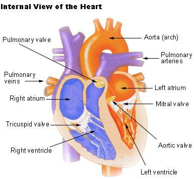
2.63: Describe the structure of the heart and how it functions
• The left and right atria are located on the top half of the heart. They receive blood from around the body.
• The left and right ventricles are located on the bottom half of the heart. They generate enough force to pump blood around the body.
• The right atrium receives deoxygenated blood through the superior and inferior vena cava. The right ventricle receives blood from the right atrium and the contraction sends the deoxygenated blood to the lungs.
• The left atrium receives oxygenated blood through the pulmonary veins.
• When the atria contract, blood from the left atrium is sent to the left ventricle.
• The left ventricle has the thickest walls of the four chambers. It receives oxygenated blood. The blood is then sent to all the tissues in the body through the aorta.
• Valves prevent the blood from going back into the heart.
2.64: Understand the heart rate changes during exercise and under the influence of adrenaline
• The heart rate increases during exercise because it has to work harder to pump oxygenated blood around the body.
• Adrenaline is a hormone produced in periods of high stress or physical activity - it causes an increase in heart rate.
• The heart rate increases during exercise because it has to work harder to pump oxygenated blood around the body.
• Adrenaline is a hormone produced in periods of high stress or physical activity - it causes an increase in heart rate.
2.65: Describe the structure of arteries, veins, and capillaries and understand their roles
• Arteries always carry blood away from the heart and they are deep under the skin. They have very thick walls and end in tiny arterioles that join into capillaries
• Veins always carry blood towards the heart and they are closer to the surface of the skin. They have thin walls and end in tiny venules that join into capillaries
• Capillaries carry blood to tissues around the body and blood is transferred from arteries to veins with capillary beds in the middle. Capillaries are the smallest blood vessels.
|
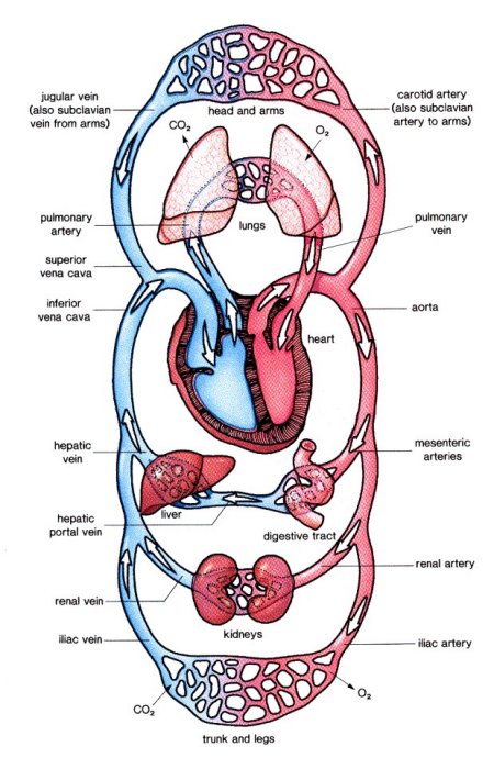
2.66: Recall the general plan of the circulation system to include blood vessels to and from the heart, the lungs, the liver, and the kidneys
|

|
i) Excretion
2.67: Recall the origin of carbon dioxide and oxygen as waste products of metabolism and their loss from the stomata of a leaf
The metabolic processes in a plant are respiration and photosynthesis:
• In photosynthesis, the oxygen is a waste product and leaves the plant via the stomata
• In respiration, the carbon dioxide is a waste product and leaves the plant via the stomata
The metabolic processes in a plant are respiration and photosynthesis:
• In photosynthesis, the oxygen is a waste product and leaves the plant via the stomata
• In respiration, the carbon dioxide is a waste product and leaves the plant via the stomata
2.68: Recall that the lungs, kidneys and skin are organs of excretion
Excretion is the removal of waste product. It is carried out by the lungs, kidneys and skin.
Excretion is the removal of waste product. It is carried out by the lungs, kidneys and skin.
- Carbon dioxide is a waste product of respiration, which is carried out by the lungs. The carbon dioxide produced during respiration is excreted by the lungs
- The kidneys excrete urine which is stored in the bladder
- The skin excretes water and salts - sweat - when the body gets too hot
|
2.69: Understand how the kidney carries out its roles of excretion and osmoregulation
|

|
2.70: Describe the structure of the urinary system, including the kidneys, ureters, bladder and urethra
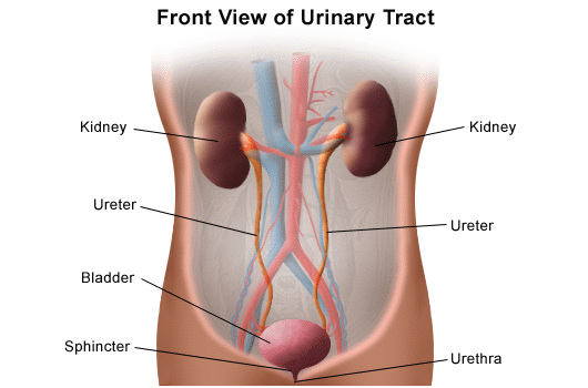
Each kidney is supplied with blood through the renal artery, which branches off the aorta, so the blood entering the kidney is at a high pressure. This blood is filtered inside each kidney and the cleaned blood passes out through the renal vein to the vena cava. The urine stored in the kidneys passes to a muscular bag called the bladder through the ureters, where it is stored. The bladder has a tube called the urethra which leads to the outside of the body. The wall of the urethra contains two ring-like muscles, called the sphincters. They can contract to close the urethra and hold back urine. The lower sphincter can be consciously controlled (people usually learn how to control it when they are toddlers), while the upper one will automatically relax when the bladder is full.
2.71: Describe the structure of a nephron, to include Bowman's capsule and glomerulus, convoluted tubules, loop of Henlé and collecting duct

click to view full size image
|
At the start of the nephron, there is a hollow cup of cells called the Bowman's capsule which is surrounded by a capillary network called the glomerulus. Part of the plasma in the blood leaves the Bowman's capsule and enters the nephron. The filtrate consists of water and small molecules. As the fluid passes along the nephron, all the glucose is absorbed back into the blood in the first coiled part of the tubule, called the proximal convoluted tubule, along with most of the sodium and chloride ions. In the rest of the tubule, more water and ions are reabsorbed, and some solutes like ammonium ions are secreted into the distal convoluted tubule. The final urine contains urea at a much higher concentration than in the blood. It also contains controlled quantities of water and ions. These quantities are controlled by the loop of Henlé, which concentrates the fluid in the tubule, causing more water to be reabsorbed into the blood, diluting it and conserving water.
|
2.72: Describe ultrafiltration in the Bowman's capsule and the composition of the glomerular filtrate
- Blood from the renal artery flows through the glomerulus - a bundle of capillaries at the start of the nephron
- A high pressure is built up which squeezes water, urea, salts, and glucose out of the blood into the Bowman's capsule (the substances which enter the Bowman's capsule can be remembered using the acronym 'HUGS', which stands for H2O, Urea, Glucose, Salts)
- The membranes between the blood vessels in the glomerulus and the Bowman's capsule act like filters, so bigger molecules like proteins and blood cells are not squeezed through and stay in the blood
- The filtered liquid in the Bowman's capsule is known as glomerular filtrate
2.73: Understand that water is reabsorbed into the blood from the collecting duct
If the body needs water for the homeostasis of blood, the water is reabsorbed back into the capillaries from the collection duct and a small volume urine is produced. It is here that the hormone ADH has an effect upon water regulation.
If the body needs water for the homeostasis of blood, the water is reabsorbed back into the capillaries from the collection duct and a small volume urine is produced. It is here that the hormone ADH has an effect upon water regulation.
2.74: Understand that selective reabsorption of glucose occurs at the proximal convoluted tubule
- All the glucose in the nephron is reabsorbed back into the blood when it reaches the proximal convoluted tubule (PCT)
- This involves active transport as it is against the concentration gradient
- Sufficient salt is reabsorbed into the blood; excess salt stays in the nephron for now
2.75: Describe the role of ADH in regulating the water content of the blood
The brain monitors the ADH levels in the blood and instructs the pituitary gland to produce more if it is needed. The loss of water from sweating produces ADH in order to increase the permeability of the collecting duct in the kidneys. This means that more water is reabsorbed back into the blood .This makes urine more concentrated so the body loses less water. This is an example of negative feedback because the change is detected and the situation is dealt with, then the corrective measure is turned off. If exercise is carried out and blood becomes too concentrated, it needs water to dilute it so ADH is released and acts in the kidneys.
The brain monitors the ADH levels in the blood and instructs the pituitary gland to produce more if it is needed. The loss of water from sweating produces ADH in order to increase the permeability of the collecting duct in the kidneys. This means that more water is reabsorbed back into the blood .This makes urine more concentrated so the body loses less water. This is an example of negative feedback because the change is detected and the situation is dealt with, then the corrective measure is turned off. If exercise is carried out and blood becomes too concentrated, it needs water to dilute it so ADH is released and acts in the kidneys.
2.76: Recall that urine contains water, urea and salts
The remaining substances in the collecting duct include water, salt and urea. These make up urine which continues out of the nephron, through the ureter and down the bladder, where it is stored before being released.
The remaining substances in the collecting duct include water, salt and urea. These make up urine which continues out of the nephron, through the ureter and down the bladder, where it is stored before being released.
j) Coordination and response
2.77: Understand that organisms are able to respond to changes in their environment
When the environment of an organism changes, they are able to respond to these changes.
When the environment of an organism changes, they are able to respond to these changes.
2.78: Understand that homeostasis is the maintenance of a constant internal environment and that body water content and body temperature are both examples of homeostasis
Homeostasis involves the balancing of bodily functions to maintain a constant internal environment. Conditions in the body need to be kept steady in order for cells to function properly. This involves the balancing of inputs and outputs.
Water enters the human body when it is drunk. It exists via urine, feces, breath, and sweat. These also have to be kept in balance - for example, when more water exits via sweat, that is, when exercise is being carried out, less water exits via urine.
Internal body temperature has to be maintained - usually at a steady 37°C. The body does this by sweating when it heats up. When it is too cold, hair on the body sticks up, providing a layer of insulation.
2.79: Understand that a coordinated response requires a stimulus, a receptor and an effector
For the body to carry out a coordinated response, several things are needed
For the body to carry out a coordinated response, several things are needed
- a stimulus - a change in the internal environment in order to trigger a response
- a receptor is needed to detect the stimulus so that it can send messages to coordinate the response
- an effector is needed to carry out the response to the stimulus
2.80: Understand that plants respond to stimuli
1. Plants increase their chances of survival by responding to changes in their environment:
2. Plants respond to predators:
3. Plants respond to abiotic (non-living) stress:
1. Plants increase their chances of survival by responding to changes in their environment:
- They sense light and grow towards it
- They sense gravity which makes the roots grow downwards and shoots grow upwards
- Climbing plants have a sense of touch in order to locate support to reach sunlight
2. Plants respond to predators:
- The white clover produces toxic chemicals which can harm predators
3. Plants respond to abiotic (non-living) stress:
- Carrots can produce antifreeze proteins at low temperatures
2.81: Describe the geotropic responses of roots and stems
Auxins are plant hormones which control growth and are produced in the tips of roots and shoots. They avoid sunlight and inhibit growth in their location. In the case of shoots, they diffuse backwards, stimulating cell growth.
Auxins are plant hormones which control growth and are produced in the tips of roots and shoots. They avoid sunlight and inhibit growth in their location. In the case of shoots, they diffuse backwards, stimulating cell growth.
2.82: Describe positive phototropism of stems
When a shoot tip is exposed to light, auxin moves to the side which is in the shade. This makes the cells grow faster on the shaded side so the shoot bends towards the light.
When a shoot tip is exposed to light, auxin moves to the side which is in the shade. This makes the cells grow faster on the shaded side so the shoot bends towards the light.
2.83: Describe how responses can be controlled by nervous or by hormonal communication and understand the differences between the two systems
- Animals increase their chances of survival by responding to changes in their environment
- They respond to changes in their internal environment to maintain their metabolism
- Any change in the environment is called a stimulus
- Stimuli are detected by receptors, which are cells found in the sense organs that can detect external stimuli
- Effectors are cells that bring about a response to stimuli, for example, glands secreting hormones
- Receptors communicate with effectors via the nervous system and/or the hormone system
|
Nervous system
|
Endocrine system
|
|
2.84: Recall that the central nervous system consists of the brain and the spinal cord and is linked to sense organs by nerves
The central nervous system (or CNS for short) consists only of the brain and spinal cord. The spinal cord is linked to receptors by nerves:
|

|
2.85: Understand that stimulation of receptors in the sense organs sends electrical impulses along nerves into and out of the central nervous system, resulting in rapid responses
The role of any receptor is to detect the stimulus by changing its energy into the electrical energy of the nerve impulses. For example, the eye converts light energy into nerve impulses, and the ear converts sound energy into nerve impulses. When energy is changed from one form to another, is is called transduction. All receptors are transducers of energy:
The role of any receptor is to detect the stimulus by changing its energy into the electrical energy of the nerve impulses. For example, the eye converts light energy into nerve impulses, and the ear converts sound energy into nerve impulses. When energy is changed from one form to another, is is called transduction. All receptors are transducers of energy:
|
Receptor
eye (sight) ear (hearing) ear (balancing) tongue (tasting) nose (smell) skin (touch/pressure/pain receptors) skin (temperature receptors) muscle (stretch receptors) |
Type of energy transduced
light sound kinetic chemical chemical kinetic heat kinetic |
2.86: Describe the structure and functioning of a simple reflex arc by the withdrawal of a finger from a hot object
A reflex is an automatic reaction, for example, the removal of your hand from a source of extreme heat. A reflex arc is the path of the reaction:
A reflex is an automatic reaction, for example, the removal of your hand from a source of extreme heat. A reflex arc is the path of the reaction:
- Receptors in the sense organ detect the stimulus (the finger detects the heat)
- Sensory neurons carry an electrical impulse to the central nervous system
- A relay neuron carries the impulse through the central nervous system where a response is decided upon
- The impulse carrying the response is sent to the effector via a motor neuron
- The effector carries out the response (the muscle contracts to bring the finger away from heat)
2.87: Describe the structure and function of the eye as a receptor
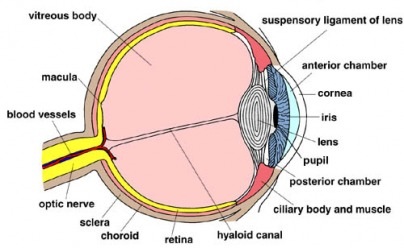
- The tough outer coat is called the sclera, the visible white part of the eye
- The transparent front of the eye is called the cornea, which lets light into the eye
- Behind the cornea, there is a colored ring of tissue called the iris
- In the middle of the iris, there is a hole called the pupil, which lets light through
- Underneath the sclera, there is a dark layer called the choroid - it stops light from being reflected around the eye
- The innermost layer of the eye is called the retina - light energy is transduced and converted to the electrical energy of nerve impulses here, and it contains rods and cones
- The rods and cones react to light, producing impulses in sensory neurons
- These sensory neurons send information to the brain via the optic nerve
- Cones, which detect color, are concentrated the the fovea
2.88: Understand the function of the eye in focusing near and distant objects, and in responding to changes in light intensity
The lens is held in place by a series of fibers called the suspensory ligaments. These are attached to a ring of muscle called the ciliary muscle. When the eye is trying to focus on a distant object, the:
When the eye is trying to focus on a nearby object, the:
The iris controls the amount of light entering the eye by changing the size of the pupil. Circular muscles form a ring in the iris, and radial muscles lie like the spokes of a wheel. When the eye is trying to focus in bright light:
When the eye is trying to focus in dim light:
The lens is held in place by a series of fibers called the suspensory ligaments. These are attached to a ring of muscle called the ciliary muscle. When the eye is trying to focus on a distant object, the:
- ciliary muscles relax
- suspensory ligaments pull tight (contract)
- lens flattens
When the eye is trying to focus on a nearby object, the:
- ciliary muscles contract
- suspensory ligaments slack (relax)
- lens is more rounded
The iris controls the amount of light entering the eye by changing the size of the pupil. Circular muscles form a ring in the iris, and radial muscles lie like the spokes of a wheel. When the eye is trying to focus in bright light:
- the circular muscles contract
- the radial muscles relax
- the pupil constricts
When the eye is trying to focus in dim light:
- the circular muscles relax
- the radial muscles contract
- the pupil dilates
2.89: Describe the role of the skin in temperature regulation, with reference to sweating, vasoconstriction and vasodilation
- When the body temperature is too hot, glands under the skin secrete sweat, which increases heat loss via evaporation
- When the body temperature is too cold, blood vessels near the surface of the skin shrink, which reduces the amount of blood that flows near the surface which means less heat is conducted by surrounding air (vasoconstriction)
- When the body temperature is too hot, blood vessels near the surface of the skin grow, which increases the amount of blood that flows near the surface which means more heat is conducted by surrounding air (vasodilation)
|
2.90: Understand the sources, roles and effects of the following hormones: ADH, adrenaline, insulin, testosterone, progesterone and estrogen
|

|
|
Gland
Pituitary Adrenals Pancreas Testes Ovaries |
Hormone
ADH (anti-diuretic hormone) Adrenaline Insulin Testosterone Estrogen Progesterone |
Function
Controls the water content of the blood Prepares the body for physical activity Lowers blood glucose Controls the development of male secondary sexual characteristics Controls development of female secondary sexual characteristics Regulates the monthly female menstrual cycle |
